Melioidosis is an irresistible sickness brought about by a Gram-contrary bacterium called Burkholderia pseudomallei. Most individuals tainted with B. pseudomallei experience no manifestations, however the individuals who do encounter side effects have signs and indications that range from gentle, for example, fever, skin changes, pneumonia, and abscesses, to extreme with irritation of the cerebrum, aggravation of the joints, and perilously low pulse that causes death. About 10% of individuals with melioidosis create indications that last more than two months, named “constant melioidosis”.
People are tainted with B. pseudomallei by contact with contaminated water. The microscopic organisms enter the body through injuries, inward breath, or ingestion. Individual to-individual or creature to-human transmission is incredibly rare. The disease is continually present in Southeast Asia, especially in upper east Thailand and northern Australia. In created nations, for example, Europe and the United States, melioidosis cases are generally imported from nations where melioidosis is more common. The signs and manifestations of melioidosis take after tuberculosis and misdiagnosis is common. Diagnosis is typically affirmed by the development of B. pseudomallei from a contaminated individual’s blood or other substantial fluid. Those with melioidosis are dealt with first with an “serious stage” course of intravenous anti-toxins (most usually ceftazidime) trailed by a multi month treatment course of co-trimoxazole. Even if the infection is appropriately treated, around 10% of individuals with melioidosis pass on from the illness. On the off chance that the infection is inappropriately treated, the passing rate could arrive at 40%.
Endeavors to forestall melioidosis incorporate wearing defensive stuff while taking care of polluted water, rehearsing hand cleanliness, drinking bubbled water, and staying away from direct contact with soil, water, or hefty rain. The anti-microbial co-trimoxazole is utilized as a preventive just for people at high danger for getting the infection in the wake of being presented to the bacteria. No antibody for melioidosis has been approved.
Approximately 165,000 individuals are tainted by melioidosis every year, coming about in around 89,000 deaths. Diabetes is a significant danger factor for melioidosis; over portion of melioidosis cases are in individuals with diabetes. Increased precipitation is related with expanded number of melioidosis cases in endemic areas. The illness was first depicted by Alfred Whitmore in 1912 in present-day Myanmar.
Sign & Symptom
Acute
The vast majority presented to B. pseudomallei experience no symptoms. About 85% of tainted individuals experience intense melioidosis. The mean hatching time of intense melioidosis is 9 days (range 1–21 days). Nevertheless, side effects of melioidosis can show up in 24 hours for those accomplished close suffocating in water. Those influenced present with manifestations of sepsis (overwhelmingly fever) with or without pneumonia, or confined sore or different focal point of contamination. The presence of vague signs and side effects has caused melioidosis to be nicknamed “the incredible mimicker”.
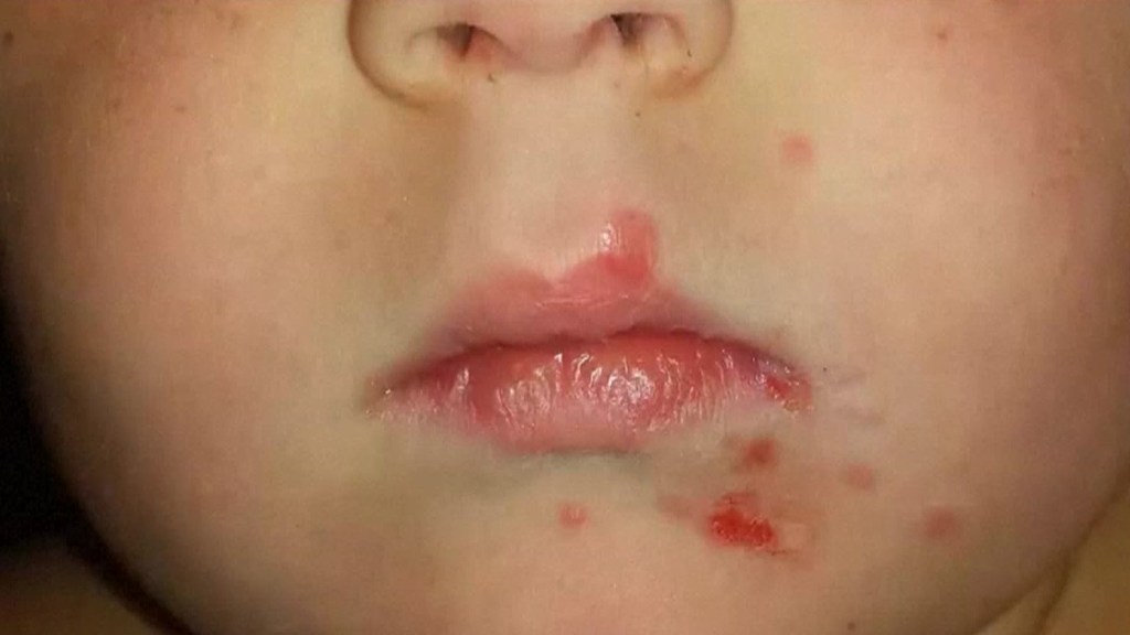
Individuals with diabetes mellitus or standard presentation to the microorganisms are at expanded danger of creating melioidosis. The illness should be considered in those remaining in endemic regions who create fever, pneumonia, or abscesses in their liver, spleen, prostate, or parotid glands. The clinical indication of the sickness can go from straightforward skin changes to serious organ problems. Skin changes can be vague abscesses or ulcerations. In northern Australia, 60% of the tainted youngsters gave just skin injuries, while 20% gave pneumonia. The commonest organs influenced are liver, spleen, lungs, prostate, and kidneys. Among the most well-known clinical signs are presence of microorganisms in blood (in 40 to 60% of cases), pneumonia (half), and septic stun (20%). People with just pneumonia may have a conspicuous hack with sputum and windedness. Be that as it may, those with septic stun along with pneumonia may have negligible coughing. Results of a chest X-beam can go from diffuse nodular invades in those with septic stun to reformist cementing of the lungs in the upper projections for those with pneumonia as it were. Overabundance liquid in the pleural depression and social occasion of discharge inside a pit are more normal for melioidosis influencing lower projections of the lungs. In 10% of cases, individuals create optional pneumonia brought about by other microorganisms after the essential infection.
Contingent upon the course of contamination, other extreme appearances create. Around 1 to 5% of those tainted create irritation of the cerebrum and mind covering or assortment of discharge in the mind; 14 to 28% create bacterial aggravation of the kidneys, kidney boil or prostatic abscesses; 0 to 30% create neck or salivary organ abscesses; 10 to 33% create liver, spleen, or paraintestinal abscesses; 4 to 14% create septic joint inflammation and osteomyelitis. Rare indications incorporate lymph hub illness taking after tuberculosis, mediastinal masses, assortment of liquid in the heart covering, unusual dilatation of veins due to infection, and aggravation of the pancreas. In Australia, up to 20% of contaminated guys create prostatic ulcer portrayed by torment during pee, trouble in passing pee, and urinary maintenance requiring catheterisation. Rectal assessment shows aggravation of the prostate. In Thailand, 30% of the tainted kids create parotid abscesses. Encephalomyelitis can happen in sound individuals without hazard factors. Those with melioidosis encephomyelitis will in general have ordinary processed tomography examines, however expanded T2 signal by attractive reverberation imaging, stretching out to the cerebrum stem and spinal line. Clinical signs include: one-sided upper engine neuron appendage shortcoming, cerebellar signs, and cranial nerve paralyses (VI, VII nerve paralyses and bulbar paralysis). A few cases gave limp loss of motion alone. In northern Australia, all melioidosis with encephalomyelitis cases had raised white cells in the cerebrospinal liquid (CSF), generally mononuclear cells with raised CSF protein.
Chronic
Constant melioidosis is typically characterized by manifestations enduring longer than two months, and happens in about 10% of patients. Clinical introductions incorporate fever, weight reduction, and profitable hack with or without grisly sputum, which may emulate tuberculosis. Furthermore, long-standing abscesses at different body destinations may likewise present. Tuberculosis should be thought of if lymph hubs are extended at the base of the lung. Pneumonia brought about by melioidosis seldom causes scarring and calcification of the lungs, dissimilar to tuberculosis.
Latent
In dormant contamination, immunocompetent individuals can clear the disease without demonstrating any indications, yet under 5% of all melioidosis cases have actuation after a time of latency. Patients with idle melioidosis might be side effect free for decades. Initially, the longest period between assumed introduction and clinical introduction was believed to be 62 years in a POW in Burma-Thailand-Malaysia.Subsequent genotyping of the bacterial confine from the Vietnam veteran, however, indicated that the disconnect might not have come from the Southeast Asia, yet from South America. This restores another report that put the longest inactivity period for melioidosis as 29 years. The potential for delayed hatching was perceived in US servicemen associated with the Vietnam War, and was alluded to as the “Vietnam time-bomb”. In Australia, the longest recorded inertness time frame is 24 years. Various comorbidities, for example, diabetes, renal disappointment, and liquor addiction can incline to reactivation of melioidosis.
Cause
Bacteria
Melioidosis is brought about by a Gram-negative, motile, saprophytic bacterium named Burkholderia pseudomallei. The microorganisms can likewise be astute, facultative intracellular pathogens. It is additionally oxygen consuming and oxidase test positive. A vacuole at the focal point of the bacterium causes it to look like a “self locking pin” when Gram stained. The microscopic organisms emanate a solid soil smell following 24 to 48 hours of development in culture. B. pseudomallei produces a glycocalyx polysaccharide container that makes it impervious to numerous sorts of antibiotics. It is commonly impervious to gentamicin and colistin, however delicate to amoxicillin/clavulanic corrosive (co-amoxiclav). B. pseudomallei is a biosafety level 3 microorganism, which requires specific research center handling. In creatures, another comparable life form named Burkholderia mallei is the causative specialist of the illness glanders. B. pseudomallei can be separated from another firmly related, yet less pathogenic species, B. thailandensis, by its capacity to acclimatize arabinose. B. pseudomallei is exceptionally versatile to different host conditions going from inside mycorrhizal organisms spores to amoebae. Its flexibility may give it an endurance advantage in the human body.
The genome of B. pseudomallei comprises of two replicons: chromosome 1 encodes housekeeping elements of the microorganisms, for example, cell divider blend, portability, and digestion; chromosome 2 encodes capacities that permit the microbes to adjust to different conditions. Even quality exchange among microscopic organisms has brought about profoundly factor genomes in B. pseudomallei. Australia has been recommended as the early store for B. pseudomallei in view of the high hereditary fluctuation of the microscopic organisms found in this district. Microorganisms separated from Africa and Central and South America appear to have a typical progenitor that lived in the seventeenth to nineteenth centuries. B. mallei is a clone of B. pseudomallei that has lost generous segments of its genome as it adjusted to live only in mammals.
Transmission
B. pseudomallei is ordinarily found in soil and surface water, and is generally bountiful at soil profundities of 10 cm to 90 cm. It has been found in soils, lakes, streams, pools, stale water, and paddy rice fields. It can make due in supplement helpless conditions, for example, refined water, desert soil, and supplement drained soil for more than 16 years. It can likewise get by in germ-free and cleanser arrangements, acidic conditions (pH 4.5 for 70 days), and conditions at temperatures going from 24 to 32 °C (72 to 89.6°F). The microscopic organisms don’t make due within the sight of bright light.
Microbes can enter the body through injuries, inward breath, and ingestion of dirtied water. Person-to-individual transmission is incredibly rare. Melioidosis is a perceived infection in creatures including felines, canines, goats, sheep, and ponies. Cows, water bison, and crocodiles are viewed as moderately impervious to melioidosis regardless of their consistent introduction to contaminated wate and soil. Flying creatures are likewise impervious to melioidosis. Transmission from creatures to people is rare.
Lacking chlorination of water gracefully had been related with B. pseudomallei episode in Northern and Western Australia. The microorganisms have additionally been found in an unchlorinated water flexibly in provincial Thailand. Water system liquid tainted with B pseudomallei is related with nosocomial injury contamination in hospitals. Based all in all genome sequencing of the microorganisms, people may assume a part in moving B. pseudomallei from spot to place.
Pathogenesis
B. pseudomallei can contaminate different kinds of cells and to sidestep human safe reactions. Microorganisms initially enter at a break in the skin or mucous layer and duplicate in the epithelial cells. From that point, they use flagellar motility to spread and contaminate different cell types. In the circulatory system, the microbes can taint the two phagocytes and nonphagocytes. B. pseudomallei utilizes flagella to move close to have cells, at that point appends to the cells utilizing different attachment proteins, including the sort IV pilus protein PilA and grip proteins BoaA and BoaB. Furthermore, bond of the microbes somewhat relies upon the presence of the host protein protease-initiated receptor-1, which is available on the outside of endothelial cells, platelets, and monocytes. When bound, the microorganisms enter have cells through endocytosis, winding up inside an endocytic vesicle. As the vesicle ferments, B. pseudomallei utilizes its sort 3 discharge framework (T3SS) to infuse effector proteins into the host cell, upsetting the vesicle and permitting the microbes to escape into the host cytoplasm. Inside the host cytoplasm, the microbes sidestep being slaughtered by the host autophagy utilizing different T3SS effector proteins. The microscopic organisms imitate in the host cytoplasm.
Inside the host cell, the microorganisms move by inciting the polymerization of the host actin behind them, moving the microscopic organisms forward. This actin-interceded motility is cultivated with the autotransporter BimA, which collaborates with actin at the last part of the bacterium. Moved by actin, the microorganisms push against the host layer, making bulges that stretch out into neighboring cells. These distensions cause neighboring cells to combine, prompting the arrangement of multinucleated monster cells (MNGCs). At the point when MNGCs lyse, they structure plaques (a focal away from with a ring of intertwined cells) that give asylum to the microbes for additional replication or inactive contamination. This equivalent cycle in tainted neurons can permit microscopic organisms to go through nerve establishes in the spinal string and cerebrum, prompting aggravation of the mind and spinal string. Other than spreading from cell to cell, the microbes can likewise spread through the circulatory system, causing sepsis. The microbes can make due in antigen-introducing cells and dendritic cells. Subsequently, these cells go about as vehicles that transport the microbes into the lymphatic framework, causing far reaching dispersal of the microscopic organisms in the human body.
While B. pseudomallei can get by in phagocytic cells, these cells can slaughter B. pseudomallei by a few instruments. Macrophages enacted by interferon gamma have improved the murdering of B. pseudomallei by means of the creation of inducible nitric oxide synthase. Fermentation of the endosome and corruption of the microscopic organisms is likewise conceivable, in any case, the bacterial container and LPS makes B. pseudomallei impervious to lysosomal debasement. When B. pseudomallei escapes into the host cytosol, it tends to be perceived by design acknowledgment receptors, for example, NOD-like receptors, setting off the development of the inflammasome and actuation of caspase 1, which instigates demise of the host cell by pyroptosis and further enactment of the safe framework. A few foundational have safeguards likewise add to the safe reaction. B. pseudomallei triggers both the supplement framework and coagulation course, anyway the thick bacterial case forestall the activity of the supplement layer assault complex.
Extra components of the insusceptible framework are initiated by the host cost like receptors, for example, TLR2, TLR4, and TLR5 that perceive the saved bits of the microorganisms, for example, LPS and flagella. This enactment brings about the creation of cytokines, for example, interleukin 1 beta (IL-1β) and interleukin 18 (IL-18). IL-18 expands IFN creation through normal executioner cells, while IL-1beta diminishes the IFN creation. These insusceptible atoms drive the enrollment of other safe cells, for example, neutrophils, dendritic cells, B cells, and T cells to the site of contamination. Lymphocytes appear to be especially significant for controlling B. pseudomallei; T cell numbers are expanded in survivors, and low T cell numbers are related with a high danger of death from melioidosis. In spite of this, HIV disease isn’t a danger factor for melioidosis. In spite of the fact that macrophages show liberated cytokine reactions in people with HIV disease, bacterial disguise and intracellular executing are as yet viable. Individuals tainted with B. pseudomallei create antibodies against the microbes, and individuals who live in endemic zones will in general have antibodies in their blood that perceive B. pseudomallei, yet the viability of these antibodies at forestalling melioidosis is unclear.
B. pseudomallei can stay inert in the human body from 19 to 29 years until it is reactivated during immunosuppression or stress response.The site of microbes during inactive disease and the component by which they dodge safe acknowledgment for quite a long time are both indistinct. Among the components proposed are dwelling in the core of the phone to forestall being processed, entering a phase of more slow development, anti-microbial opposition, and hereditary adaption to the host climate. Granulomas (containing neutrophils, macrophages, lymphocytes, and multinucleated goliath cells) framed at the contamination site in melioidosis have been related with inert disease in humans.
Diagnosis
Bacterial culture is the complete analysis of melioidosis. B. pseudomallei is never important for human verdure. Consequently, any development of the microorganisms is demonstrative of melioidosis. Blood societies are the most widely recognized examples for conclusion, as microbes can be identified in the blood in 50 to 60% of melioidosis cases. Different examples, for example, throat, rectal swabs, discharge from abscesses, and sputum, can likewise be utilized for culture. At the point when microbes don’t develop from individuals emphatically associated with having melioidosis, rehashed societies should be taken, as ensuing societies can become positive. B. pseudomallei can be developed on sheep blood agar, MacConkey agar, Ashdown’s medium (containing gentamicin), or Ashdown’s stock (containing colistin). Agar plates for melioidosis should be hatched at 37°C (98.6°F) in air and reviewed day by day for four days. On the agar plates, B. pseudomallei structures velvety, nonhaemolytic, provinces following 2 days of brooding. Following 4 days of brooding, states seem dry and wrinkled. Colonies of B. pseudomallei that are become on Francis medium (an adjustment of Ashdown medium with gentamicin focus expanded to 8 mg/l) are yellow. For research facilities situated external endemic zones, Burkholderia cepacia particular agar or Pseudomonas specific agar can be utilized if Ashdown’s medium isn’t available. It is significant not confound the bacterial development as Pseudomonas or Bacillus spp. Other biochemical screening apparatuses can likewise be utilized for identifying B. pseudomallei, including the API 20NE or 20E biochemical pack joined with Gram stain, oxidase test, common development qualities, and protection from specific anti-microbials of the bacteria. Molecular techniques, for example, 16S rDNA tests and polymerase chain response can likewise be utilized to distinguish B. pseudomallei in culture, yet they are just accessible in exploration and reference laboratories.
General blood tests in individuals with melioidosis show low white platelet tallies (demonstrates contamination), raised liver compounds, expanded bilirubin levels (demonstrates liver brokenness), and raised urea and creatinine levels (demonstrates kidney brokenness). Low blood glucose and acidosis predicts a less fortunate visualization in those with melioidosis. Nonetheless, different tests, for example, C-receptive protein and procalcitonin levels are not solid in anticipating the seriousness of melioidosis infection.
By microscopy, B. pseudomallei is viewed as Gram-negative and pole molded, with a bipolar recoloring comparable in appearance to a self clasping pin. Microbes can in some cases be seen straightforwardly in clinical examples from contaminated individuals, yet ID by light microscopy is neither explicit nor touchy. Immunofluorescence microscopy is profoundly explicit for recognizing microbes straightforwardly from clinical examples, yet has under half sensitivity. A parallel stream immunoassay has been grown however not broadly evaluated. An expanding number of labs use Matrix-helped laser desorption/ionization mass spectrometry to distinguish the microorganisms accurately.
Serological tests, for example, circuitous haemagglutination have been utilized to identify the presence of antibodies against B. pseudomallei. Various gatherings of individuals, however, have generally various degrees of antibodies, so understanding of these tests relies upon area. In Australia, under 5% of individuals have B. pseudomallei antibodies, so the presence of even moderately low measures of immune response is irregular and could propose melioidosis. In Thailand, numerous individuals have antibodies against B. pseudomallei, so just a moderately high measure of immunizer in the blood proposes melioidosis. Thailand likewise utilizes direct immunofluorescent neutralizer test (IFAT) and latex agglutination. In IFAT, both B. pseudomallei antigen and B. thailandensis can be utilized to measure the measure of antibodies delivered against the microscopic organisms. In this way, the outcomes must be deciphered with alert as a bogus positive response could be found on the off chance that somebody is recently presented to nonpathogenic B. thailandensis. Latex agglutination is valuable in screening for suspected B. pseudomallei colonies. Commercial ELISA packs for melioidosis are not, at this point accessible in the market because of low affectability to human antibodies detection.
Different imaging modalities can likewise help with the conclusion of melioidosis. In intense melioidosis with the spreading of the microbes through the circulatory system, the chest X-beam shows multifocal nodular injuries. It might likewise show consolidating knobs or cavitations. For those with intense melioidosis without the spread to the circulation system, chest X-beam shows upper-flap union or cavitations. In persistent melioidosis, the moderate movement of upper-flap solidification of the lungs takes after tuberculosis. For abscesses situated in different pieces of the body separated from the lungs, particularly in the liver and spleen, CT examine has higher affectability when contrasted and a ultrasound check. In liver and splenic abscesses, a ultrasound check shows “target-like” sores, while CT filter shows “honeycomb sign” in liver abscesses. For melioidosis including the mind, MRI has higher affectability than a CT examine in diagnosing the injury. X-ray shows ring-improving sores for mind melioidosis.
Prevention
Melioidosis is a notifiable infection in Australia. This empowers the nation to screen illness trouble and contain episodes. Melioidosis has just been a notifiable condition in Thailand since June 2016. All things considered, Australia additionally set out on mindfulness missions to expand the network’s comprehension of the disease. In the United States, lab laborers can deal with clinical examples of B. pseudomallei under BSL-2 conditions, while large scale manufacturing of such creatures requires BSL-3 precautions. Also, a few instances of medical clinic gained disease of melioidosis have been accounted for, so medical care suppliers are prescribed to rehearse hand cleanliness and widespread precautions.
Huge scope water chlorination has been fruitful at diminishing B. pseudomallei in the water in Australia. In center to low-pay nations, water should be bubbled before utilization. In big league salary nations, water could be treated with bright light for those in danger of contracting melioidosis. The individuals who are at high danger of contact with the microorganisms should wear defensive stuff, (for example, boots and gloves) during work. Those remaining in endemic territories ought to stay away from direct contact with soil, and open air introduction to hefty downpour or residue mists. Filtered water or bubbled water are favored for drinking.
Postexposure prophylaxis
After introduction to B. pseudomallei (especially following a research center mishap), treatment with co-trimoxazole is suggested. Then again, co-amoxiclav and doxycycline can be utilized for the individuals who are narrow minded to co-trimoxazole. Since co-trimoxazole can cause extreme results, just high-hazard people will in general get such medicines. Okay people would get incessant observing, instead.
Vaccination
A few antibody applicants have been tried in creature models. By the by, no antibody up-and-comers have been attempted in people. Significant obstacles of the antibodies are restricted adequacy in creature models, setting up the best technique for immunization organization in people, and calculated and monetary issues in building up human preliminaries in endemic areas.
Treatment
The therapy of melioidosis is isolated into two phases, an intravenous escalated stage and a destruction stage to forestall repeat. The selection of anti-toxins relies on the helplessness of the microscopic organisms to different anti-infection agents. B. pesudomallei is commonly powerless to ceftazidime, meropenem, imipenem, and co-amoxiclav. These medications are intended to slaughter the microorganisms. It is likewise defenseless to doyxcycline, chloramphenicol, and co-trimoxazole. These medications are intended to hinder the development of the microscopic organisms. The microscopic organisms are impervious to penicillin, ampicillin, first-and second-age cephalosporin, gentamicin, streptomycin, tobramycin, macrolides, and polymyxins. B. pseudomallei segregates from the locale of Sarawak, Malaysia are helpless to gentamicin, though.
Intensive phase
Intravenous ceftazidime is the current medication of decision for treatment of intense melioidosis and should be controlled for at any rate 10 days. Meropenem, imipenem, and the cefoperazone-sulbactam mix (Sulperazone) are additionally effective. Intravenous amoxicillin-clavulanate (co-amoxiclav) might be utilized if nothing unless there are other options four medications is available; co-amoxiclav keeps passing from melioidosis, as does ceftazidime. Intravenous anti-microbials are given for at least 10 days. The middle fever freedom time in melioidosis is 9 days.
Meropenem is the favored anti-infection treatment for neurological melioidosis and those with septic stun conceded into concentrated consideration units. Co-trimoxazole is suggested for neurological melidosis, osteomyelitis, septic joint pain, skin and gastrointestinal disease, and profoundly situated boil. For profound situated contaminations, for example, abscesses of interior organs, osteomyelitis, septic joint pain, and neurological melioidosis, the length of anti-microbials given should be longer (dependent upon 4 to about two months). The time taken for fever to be settled can be over 10 days in those with profound situated contamination. Protection from ceftazidime, carbapenems, and co-amoxiclav are uncommon in the concentrated stage, however are more noticeable during annihilation treatment. No distinctions are seen between utilizing cefoperazone/sulbactam or ceftazidime to treat melioidosis, as both show comparative demise rates and illness movement following treatment. For those with kidney weakness, the measurement of ceftazidime, meropenem, and co-trimoxazole should be lowered. Once the clinical condition improved, meropenem can be exchanged back to ceftazidime. Whether the ceftazidime or meropenem mix treatment decreases backslide rates in beginning stage of the treatment is unclear.
Eradication phase
In Australia, co-trimoxazole is utilized in kids and pregnant moms after the initial 12 weeks of pregnancy. Then, in Thailand, co-amoxiclav is the medication of decision for kids and pregnant ladies. Notwithstanding, B. pseudomallei regularly gets obstruction when co-amoxiclav is utilized. Cases have additionally been accounted for where melioidosis is effectively treated with co-trimoxazole for a very long time without experiencing concentrated treatment gave that lone skin indications are seen without the association of inside organs or sepsis. Resistance to cotrimoxazole is uncommon in Australia.
Surgery
Careful seepage is shown for single, huge abscesses in the liver, muscle, and prostate. Notwithstanding, for various abscesses in the liver, spleen, and kidney, careful seepage may not be conceivable or important. For septic joint inflammation, arthrotomy waste of time and seepage is required. Careful debridement might be fundamental. For those with mycotic aneurysm, dire medical procedure is needed for prosthetic vascular unions. Deep rooted treatment with co-trimoxazole might be required for those with prosthetic vascular unions. Different abscesses infrequently should be depleted on the grounds that most of them can resolve with anti-infection treatment. In Australia, prostate canker may require routine imaging and drainage.
Others
Immunomodulating treatments, for example, granulocyte state invigorating factor, Interleukin 7, and against PDI (customized cell passing) could be valuable in melioidosis treatment, particularly for those with septic stun. This is on the grounds that these medications could assist with boosting the human body safe capacity against the bacteria.
Prognosis
In well-resourced settings, where the infection can be identified and treated early, the danger of death is 10%. In asset helpless settings, the danger of death from the sickness is more than 40%.
For those with deficient treatment, return of indications after a time of infection reduction (“recrudescence”) can happen. At that point, clinic affirmation is required for intravenous anti-microbials. For the individuals who have finished treatment effectively, repeat can likewise happen because of recrudescence or new melioidosis contamination. With better treatments, the recrudescence rate has diminished from 10 to 5%. The new contamination is currently more normal than recrudescence. Danger components of recrudescence incorporate the seriousness of infection (patients with positive blood societies or multifocal illness have a higher danger of backslide), decision of anti-infection for annihilation treatment (doxycycline monotherapy and fluoroquinolone treatment are not as viable), helpless consistence with destruction treatment and term of annihilation treatment under 8 weeks.
Fundamental ailments, for example, diabetes mellitus, ongoing kidney sickness, and malignant growth can decline the drawn out endurance and inability of the individuals who recuperate from disease. The most serious entanglement of melioidosis is encephalomyelitis. It can cause quadriparesis (muscle shortcoming in all the appendages), incomplete limp paraparesis (muscle shortcoming of the two legs), or foot drop. For those with past melioidosis-related bone and joint contaminations, inconveniences, for example, sinus disease, bone and joint deformations with restricted scope of movement can occur.
Epidemiology
Melioidosis is an understudied infection that stays endemic in non-industrial nations. In 2015, the International Melioidosis Society was shaped to bring issues to light of the disease. In 2016, a factual model was created which demonstrated that the number is 165,000 cases for each year with 138,000 of those happening in East and South Asia and the Pacific. In about portion of those cases (54% or 89,000), individuals will die. Under-detailing is a typical issue as just 1,300 cases were accounted for worldwide since 2010, which is under 1% of the extended occurrence dependent on the modeling. Lack of lab symptomatic abilities and absence of infection mindfulness among medical services suppliers additionally causes underdiagnosis. Regardless of whether bacterial societies turn positive for B. pesudomallei, they can be disposed of as pollutants particularly in labs in non-endemic areas. As of 2018, melioidosis is excluded from the WHO rundown of dismissed tropical diseases.
Melioidosis is endemic in pieces of Southeast Asia (counting Thailand, Laos, Singapore, Brunei, Malaysia, Myanmar and Vietnam), southern China, Taiwan and northern Australia. Heavy precipitation can expand its degree into focal Australia. India, and irregular cases in South America. The genuine weight of melioidosis in Africa and Middle East stay obscure because of low measure of information. There were 24 African nations and three Middle Eastern nations anticipated to be endemic with melioidosis, anyway not a solitary case was accounted for from them. An aggregate of 51 instances of melioidosis were accounted for in Bangladesh from 1961–2017. Regardless, absence of mindfulness and assets offers ascend to underdiagnosis of the infection in the country. In the United States, two verifiable cases (1950 and 1971) and three ongoing cases (2010, 2011, 2013) have been accounted for among individuals that didn’t travel abroad. Regardless of broad examinations, the wellspring of melioidosis was rarely affirmed. One potential clarification is that importation of therapeutic plant items or fascinating reptiles might have brought about the presentation of melioidosis in the United States. In Europe, the greater part of the melioidosis cases are imported from Thailand.
Melioidosis is found in all age groups. For Australia and Thailand, the middle time of contamination is at 50 years; 5 to 10% of the patients are under 15 years. The absolute most significant danger factor for creating melioidosis is diabetes mellitus, trailed by risky liquor use, persistent kidney illness, and constant lung disease. Greater than half of individuals with melioidosis have diabetes; diabetics have a 12-overlap expanded danger of contracting melioidosis. Diabetes diminishes the capacity of macrophages to battle the microbes and lessens T aide cell creation. Inordinate arrival of tumor rot factor alpha and interleukin 12 by mononuclear cells builds the danger of septic stun. The diabetes drug glibenclamide can likewise dull monocyte’s provocative responses. Other danger factors incorporate thalassaemia, word related presentation (for example rice paddy farmers), recreational presentation to soil, water, being male, age more noteworthy than 45 years, and delayed steroid use/immunosuppression, yet 8% of youngsters and 20% of grown-ups with melioidosis have no danger factors. HIV contamination doesn’t incline to melioidosis. Infant cases have been accounted for potentially because of mother-to-kid transmission, network gained disease, or medical care related infection. Those who are well may likewise be tainted with B. pseudomallei. For instance, 25% of kids remaining in endemic regions began delivering antibodies against B. pseudomallei between a half year and 4 years of age, recommending they were presented to it throughout this time. This implies that numerous individuals without side effects will test positive in serology tests in endemic areas. In Thailand, the seropositivity rate surpasses half, while in Australia, the seropositivity rate is just 5%. The infection is obviously connected with expanded precipitation, with the quantity of cases rising after expanded precipitation. Extreme precipitation expands the centralization of the microscopic organisms in the dirt, accordingly expanding the danger of communicating the microorganisms through the air.
History
Pathologist Alfred Whitmore and his collaborator Krishnaswami first revealed melioidosis among bums and morphine addicts at post-mortem examination in Rangoon, present-day Myanmar, in a report distributed in 1912. Arthur Conan Doyle may have perused the 1912 report prior to composing a short story that elaborate the invented tropical sickness “Tapanuli fever” in a Sherlock Holmes adventure. In the 1913 story of “The Dying Detective”, Holmes got a crate intended to immunize the casualty with “Tapanuli fever” after opening. “Tapanuli fever” was thought by numerous individuals to speak to melioidosis. The expression “melioidosis” was first instituted in 1921. It was recognized from glanders, an infection of people and creatures that is comparative in introduction, however brought about by an alternate miniature living being. B. pseudomallei, otherwise called the Whitmore bacillus, was distinguished in 1917 in Kuala Lumpur. The primary human instance of melioidosis was accounted for in Sri Lanka in 1927. In 1932, 83 cases were accounted for in South and Southeast Asia with 98% mortality. In 1936, the main creature (sheep) instance of melioidosis was accounted for in Madagascar, South Africa. In 1937, soil and water were recognized as the environments of B. pseudomallei. During the Vietnam War from 1967 to 1973, 343 American officers were accounted for with melioidosis, with around 50 cases sent through inhalation. A flare-up of melioidosis at the Paris Zoo during the 1970s (known as L’affaire du jardin des plantes) was thought to have started from an imported panda or ponies from Iran. The primary proof of B. pseudomallei (in soil) in Brazil was accounted for in 1983.
Preceding 1989, the standard treatment for intense melioidosis was a three-drug blend of chloramphenicol, co-trimoxazole, and doxycycline; this routine is related with a death pace of 80% and is not, at this point utilized except if no different choices are available. All three medications are bacteriostatic (they prevent the bacterium from developing, however don’t murder it) and the activity of co-trimoxazole irritates both chloramphenicol and doxycycline. Aerosolised B. pseudomallei was first segregated in 1989. In the exact year, ceftazidime had been appeared to diminish the danger of death of melioidosis from 74% to 37%. B. pseudomallei was recently delegated part of the family Pseudomonas until 1992. In 1992, the microbe was officially named B. pseudomallei. The name melioidosis is gotten from the Greek melis (μηλις) signifying “a sickness of asses” with the postfixes – oid signifying “like” and – osis signifying “a condition”, that is, a condition like glanders. In 2002, B. pseudomallei was named a “class B specialist”. A live constricted immunization was created in mice in the very year. In 2003, multilocus arrangement composing for B. pseudomallei was created. In 2012, B pseudomallei was delegated a “level 1 select specialist” by the U.S. Habitats for Disease Control. In 2014, co-trimoxazole was set up as the oral destruction treatment. In 2015, B. pseudomallei DNA was distinguished in separated air utilizing quantitative PCR. In 2016, a factual model was created to anticipate the event of worldwide melioidosis every year. In 2017, entire genome sequencing recommended Australia as the early repository for melioidosis.